
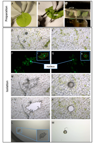
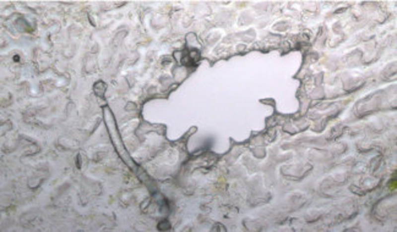
Laser dissection microscopy (LDM) is a method for obtaining specific tissues, cells, or even organelles under direct microscopic observation to enrich histologically nearly pure cell populations. In the recent publication, a method for amplifying viral circular DNA from single nuclei or cells by rolling circle amplification (RCA) is presented to show and proof infection of the selected and collected nucleus, nuclei or cell(s), respectively. For this purpose, our group used a MMi Cellcut microdissection system. In addition, proof of principle was provided that quantification of viral DNA molecules in a single plant nucleus is possible. Furthermore, a method is described to isolated non-fixed epidermal tissue from Nicotiana benthamiana plants/leaves and Arabidopsis thaliana by peeling suitable for the preparation of samples for LDM.
In the future, the results will enable quantitative analyses at the single cell level as well as evolutionary analyses, e.g. to determine quasi-viral species in a single cell. Of particular note is that the combination of RCA or PCR analyses of LDM-based dissected organelles is not limited to a plant pathogen/host system and can be transferred to other systems.
Original publication
Sicking C, Krenz B. (2022) Rolling circle amplification of begomoviral DNA from a single nucleus isolated by laser dissection microscopy. J Virol Methods. 2022 Oct;308:114591.
doi: 10.1016/j.jviromet.2022.114591. Epub 2022 Jul 23