We focus on two major types of disease models in our group:
1. 2D/3D muscle disease models
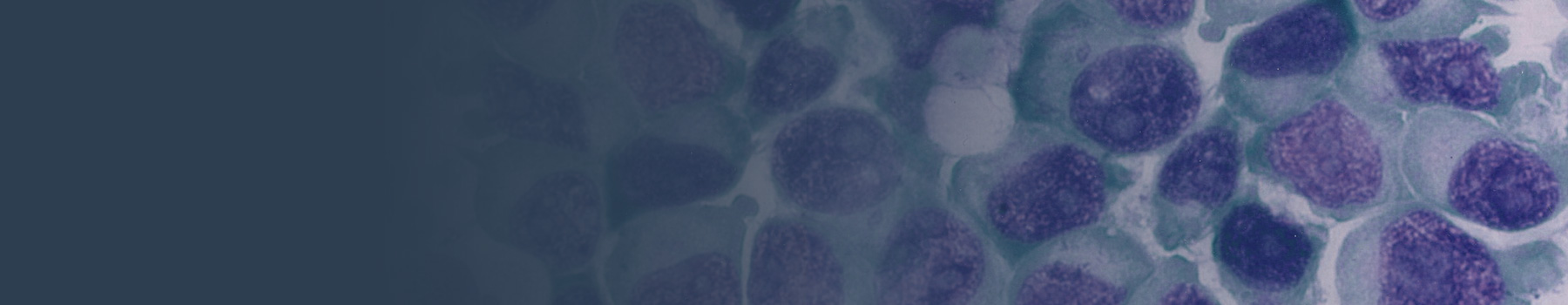
The muscle stem cells or myoblasts are the valuable source to model muscle diseases due to their remarkable ability to regenerate new myofibers. The primary muscle cells are significantly limited in applications by their inability to expand in culture for long term. The immortalized myoblasts or iPSCs are becoming alternative, in particular the patient-derived iPSCs due to its characteristics such as immortality, multi-lineage differentiation potential to 3D muscles, and patient genomic specificity. The CRIPSR tools can either be used to correct mutations in the disease models or generate mutation carrying cell lines to model muscle diseases.

Project: CNMuscle, funded by ZNM - Zusammen Stark! e.V.
Duration: 01.11.2023 - 30.04.2026
More information can be found here.
2. 2D Neuronal disease models
For the NMDs affecting the nerves or brain, the in vitro neuronal model allows the systematic evaluation of the mutated neuronal receptors or transporters, further providing a genotype-phenotype correlation or suggesting treatment strategies.

If you are interested in our disease cell models, please contact us and we are glad to collaborate. Students and scientists are also welcome to join our team.
Selected References
- Wang H#, Krause A, Escobar H, Müthel S, Metzler E, Spuler S#. LMNA Co-Regulated Gene Expression as a Suitable Readout after Precise Gene Correction. International Journal of Molecular Sciences 2022; 23 (24), 15525. (Co-corresponding authors).
- Wang H, Kaçar Bayram A, Sprute R, Ozdemir O, Cooper E, Pergande M, ..., Cirak S. Genotype-Phenotype Correlations in Charcot-Marie-Tooth Disease Due to MTMR2 Mutations and Implications in Membrane Trafficking. Frontiers in Neuroscience 2019; 13, 974.
- Wang H*, Schänzer A*, Kampschulte B, Daimagüler HS, Logeswaran T, Schlierbach H, ..., Cirak S. A novel SPEG mutation causes non-compaction cardiomyopathy and neuropathy in a floppy infant with centronuclear myopathy. Acta Neuropathologica Communications 2018; 6 (1), 1-5. (Co-first authors)
- Wang H*, Salter CG*, Refai O*, Hardy H, Barwick KES, Akpulat U, ..., Crosby AH. Choline transporter mutations in severe congenital myasthenic syndrome disrupt transporter localization. Brain 2017; 140 (11), 2838-2850. (Co-first authors)
- Baarlink C, Wang H, Grosse R. Nuclear actin network assembly by formins regulates the SRF coactivator MAL. Science 2013; 340 (6134), 864-867.